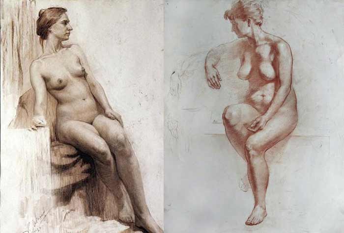
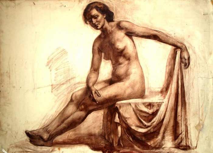
How to Draw Female Body
How to Draw Female Body
Anatomy Lesson 15 – Part 2
In this video lesson, you will discover how to draw female body.
How to Draw Female Body for Figurative Artists
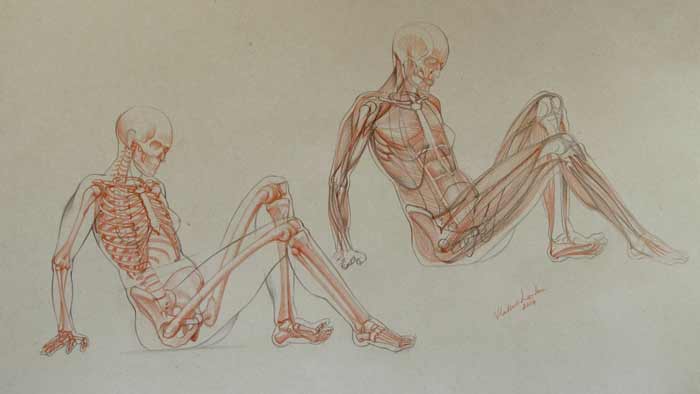
When it comes to the topic of how to draw female body, a figurative artist should know the muscle anatomy.
Let’s begin with the description of major muscles.
The deltoid muscle has a triangular shape. This powerful muscle starts from the outer half of the collarbone and from the shoulder blade and inserts into the upper arm bone. Its main action is to lift the arm sideways and in front of the torso. This muscle also contributes to extension of the arm, moving it backward behind the torso.
Another powerful muscle of the upper body is the chest muscle. It inserts into the upper art bone underneath the deltoid muscle.
The chest muscle consists of three parts:
- The abdominal portion goes from the sixth and seventh ribs to the top point of insertion.
- The middle portion goes from the breastbone and inserts into the middle point of insertion.
- And the top part of this muscle goes from the inner half of the collarbone and inserts into the lower point of insertion.
The main function of the chest muscle is to move the upper arm in adduction, flexion, and medial rotation. Three portions of this muscle cross each other in the armpit area.
Now, let’s check the prominent muscle of the neck.
It consists of two portions. The front one travels from the top of the breastbone and the second portion is connected to the upper inner half of the collarbone. These two portions insert into the mastoid process of the skull, which is just behind the ear.
This neck muscle is covered by another muscle, which is wide and flat and travels from the collarbone to the lower jaw. This muscle covers the front side of the neck and the under-plane of the chin.
The upper portion of the trapezius muscle covers the back side of the neck.
The major flexor of the arm is the biceps brachii. It occupies the entire front portion of the upper arm. This muscle has two heads. One portion goes from the coracoid process of the shoulder blade and another from the point nearby, the arm joint. The tendon of this portion travels through the groove at the top of the upper arm bone. At its lower end, the biceps brachii inserts into the upper sections of two bones of the forearm. When contracted, it flexes the arm at the elbow joint.
Underneath this muscle, there is another one, which begins from the middle part of the upper arm bone and inserts into the elbow bone. It helps flex the arm.
The main extensor of the arm is the triceps brachii. It covers the most rear area of the upper arm. As indicated by its name, this muscle has three heads. Two heads are attached to the upper arm bone and the third one goes from the shoulder blade. All three heads are inserted, via the common tendon, into the elbow.
The right arm is depicted in the pronation position. This is when the radius bone is rotated around the elbow bone. In such position, the extensor muscles are rotated together with the radius. They travel from the lower end of the upper arm and insert into various bones of the hand.
When drawing the lower arm, you need to remember that the most bulk of these muscles is located in the upper half of the lower arm.
Approximately, midway down, these muscles become narrow tendons, which makes the arm slimmer.
Here is the fact to remember when drawing the arm: the extensor muscles travel from the outer epicondyle of the upper arm bone. In opposition to the extensors, the flexor muscles of the arm travel from the inner epicondyle of the upper arm bone. The main function of flexor group of muscles is to flex the hand and fingers.
The six-pack muscle inserts into the ribcage at the fifth, sixth, and seventh coastal cartilages of ribs, and also attaches to the lower part of the breastbone. This long muscle starts from the pubic bone and covers the entire abdominal portion of the body. In the middle, it is separated into two vertical bands by the white line. Horizontally, three fibrous lines divide this muscle, which together with the white line splits this muscle into six-packs. Such division allows separate movements of this muscle.
At the side of the six-pack muscle, there is the white line, which is called the semi-lunar line. Next to this line, is the internal oblique muscle; it wraps the body from both sides. This internal muscle has two primary functions. It acts in opposition to the diaphragm, reducing the volume of the chest when a person exhales. It also rotates and side bends the torso by pulling the ribcage toward the hip and lower back.
The muscle, which looks like ribs, is called the serratus anterior. It begins from the sides of the first eight or nine ribs and inserts into the inner border of the shoulder blade underneath this bone. The lower four digitations of this muscle intervene with the external oblique muscle which covers the frontal and side part of the abdomen.
The external oblique muscle begins from the outer surface of the lower eight ribs (ribs five through twelve). It not only rotates the torso, it also bends it forward and sideways.
There is one muscle of the back that inserts into the upper arm. It originates from the spinal column, lower ribs, and pelvis. It is called the latissimus dorsi. It is the widest muscle of the back.
The gluteus medius muscle travels from the top ridge of the pelvis to the upper projection of the thighbone. Its main action is to raise the upper leg sideways.
Another muscle of the gluteal group is the gluteus maximus. The buttocks muscle begins from the backside of the pelvis and inserts into the upper leg bone. This muscle moves the thigh backward and also assists in rotating the upper leg outward. The buttocks muscles are activated when a person climbs upstairs or walks uphill.
From the bony projection of the frontal edge of the pelvis begins a powerful muscle that covers most of the frontal portion of the thigh. This muscle is called the quadriceps. It has four heads. Each of them is inserted, via the common tendon, into the tibial tuberosity on the shinbone. All heads of the quadriceps come together and insert into the kneecap, encapsulating it from the front and back. Thereafter, this tendon goes downward and attaches to the shinbone. The top portion of the quadriceps begins from the pelvis and other three heads originate from the top part of the thighbone. The main function of this muscle is to extend the lower leg by straightening it at the knee.
The tailor’s muscle begins from the frontal top edge of the pelvis and wraps around the thigh on the front. It travels diagonally downward to the knee area and inserts into the inner top end of the shinbone.
At the back of the thigh, there is the hamstring group of muscles. One muscle of this group is called the biceps femoris. It has two heads. One begins from the sitting bone and another starts from the shaft of the thighbone. They both insert, via the common tendon, into the top part of the calfbone.
There are two more muscles of the hamstring group. They also begin from the sitting bone. One of the muscles attaches to the inner top-side of the shinbone and another also inserts into the shinbone on its outer-top-side.
All hamstring muscles are involved in extension of the thigh and flexion of the lower leg.
Now let’s examine the muscles of the lower leg.
At the back of the lower leg, there are two major muscles – the soleus and the calf muscle.
The soleus muscle begins from the top back surfaces of the shin and calf bones.
The calf muscle has two heads, which begin from the condyles of the thighbone.
Both muscles, the soleus and the calf muscle insert, via the common tendon, into the heel bone. These muscles flex the lower leg and move the foot down.
At the outer side of the calf bone, there are two muscles that begin from the calf bone and insert into various places on the foot. They help move the foot downward and turn it outward.
At the front of the lower leg, there is an extensor muscle that begins from the top part of the upper leg and inserts into the phalanges of the lesser toes. Its primary functions are to extend the foot, move it upward, and lift the toes.
Next to this muscle, there is the shin muscle. As its name suggests, it begins from the shinbone. It inserts into the foot on its inner side. It has the function of lifting the front part of the foot upward.
In the foot, there are a couple muscles worth mentioning. Beginning from the heel bone, one muscle inserts into the phalange of the great toe and another muscle inserts into the phalanges of the lesser toes. These muscles flex the toes downward…
[ The full lesson is avaibale to Anatomy Master Class members ]
To learn more about how to draw female body, enroll in the Anatomy Master Class
Simple Pricing, No Surprises
One-time payment - Only $97 USD
ENROLL NOW