Female Anatomy for Artists
Female Anatomy for Artists
Anatomy Lesson 15 – Part 1
In this video lesson, you will discover the female anatomy for artists.
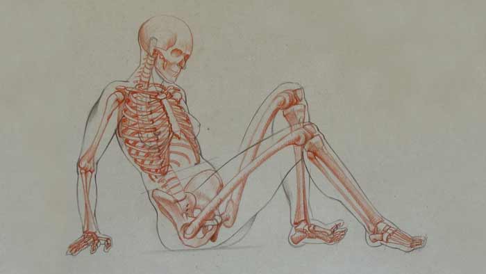
This video part will show the skeletal anatomy of a sitting female figure.
Female Anatomy for Figurative Artists
The spinal column is curved into the characteristic “S” shape. It has four regions – the regions of the neck, ribcage, waist, and sacrum. Each region is curved in the opposite direction as the previous one.
At the top of the spine, based on the first vertebra, there is the skull. All bones of the skull, apart the lower jaw, are fused together and are immovable in relation to each other.
The skull has a complicated bony structure which consists of twenty-two bones (excluding teeth).
The cranium section of the skull protects the most vital organ in the human body – the brain. This section consists of eight bones.
The zygomatic bone, or the cheek bone, is quite an important landmark on the skull.
The inner ear hole is located just below this bone.
The first pair of the ribs goes from the eighth vertebra of the spinal column. The first ribs form an oval. The diameter of this oval is equal to the width of the neck at its base. Many neck muscles are attached to this pair of ribs.
Seven vertebrae above form the neck region of the spine.
At the front, the first ribs are connected to the manubrium of sternum which is the top part of the breastbone. The length of the breastbone is equal to the height of the face.
Collarbones are connected to the top edge of the breastbone just above the first pair of ribs. The length of the collarbone is equal to the height of the face and to the length of the breastbone.
There are twelve vertebrae in the ribcage region of the spine. In the waist region, there are five vertebrae. Every following vertebra needs to be slightly larger than the previous one.
The outline of the ribcage starts from the first pair of ribs at its narrowest point. It becomes wider as it goes down with its widest point at the eighth pair of ribs.
The first seven ribs are attached to the breastbone. They are called the true ribs.
Ribs connect to the breastbone via the coastal cartilage. This cartilage is more flexible than the bony structure of the rib.
The first pair of ribs is attached in such a way that it travels upward from the breastbone.
The second pair of ribs travels sideways from the breastbone. At the back, it attaches to the second vertebra of the ribcage region of the spinal column; and at the front, it attaches to the place where the manubrium of sternum connects to the body of sternum.
Starting from the third pair of ribs and onward, the direction of attachment to the breastbone goes downward. The lower the rib on the breastbone, the larger the angle at which it goes down.
This structure allows certain movements of the ribcage. For example, when a person breathes in and out, the ribcage enlarges and shrinks.
The eighth, ninth, and tenth ribs are not directly attached to the breastbone, but instead, via coastal cartilage, they attached to the previous rib. This is why these pairs of ribs are called false ribs.
There are two more pairs, the eleventh and the twelfth. These ribs are only attached to the spinal column and at the front, they float free and do not attach to previous ribs nor the breastbone. This is why they are called floating ribs.
Both males and females have twelve pairs of ribs. The biblical story that males have one less rib is not anatomically accurate. However, there are some rare cases where a person can have more or less ribs, but this is not the rule, but an exclusion.
The distance between the pelvis and the ribcage is not so great. It is about only a couple inches.
The axis of the hip joints is on the same level as the pubic bone. This axis also runs parallel to the top edge of the pelvis and the bottom edge of the ribcage.
The vertebrae of the pelvis region form the sacrum. These verebrae are fully fused together when a person is thirty-four years of age.
The top edge of the iliac bone is called the iliac crest.
The pelvis is slightly wider than the ribcage in both males and females.
At the bottom part of the pelvis, there are two bagel-shaped bones which we will refer to by the common name – the sitting bones.
The upper arm bone is connected to the body via the shoulder blade.
The bony projection of the shoulder blade, which connects to the collarbone, is called the acromion.
The shoulder joint is a ball-and-socket type of joint.
Near the shoulder joint, there is the bony projection of the shoulder blade which resembles a bird’s beak. It is called the coracoid process.
The upper arm bone is called the humerus.
At the top, it has a round head, which is about the size of a golf ball, as well as two bony projections and a groove between them.
At the lower end of this bone, there are two projections which are the inner and outer epicondyles.
These two projections are very close to the surface of the arm and you can easily detect them under your fingers when touching the elbow from both sides.
The lower arm has two bones – the elbow bone and the radius.
In our case, the arm is in the pronation position. In this position, the radius is rotated around the elbow bone and is no longer parallel to it, but crosses above.
The thigh bone is slightly bent forward. At its lower end, it is wider and has two bony projections which are the inner and outer condyles.
The lower leg has two bones, the tibia (the shin bone) and the fibula (the calf bone).
The top end of the shinbone, together with the condyles of the thighbone, form the knee joint. This is the hinge-type joint, which allows rotation of the lower leg in relation to the upper leg.
In the front, the triangular-shaped knee cap protects the knee joint, especially when the leg is flexed.
The calf-bone is connected to the shinbone on its outer side. It is much more slender than the shinbone. Its lower end is the outer ankle bone which is positioned lower than the inner ankle.
The foot consists of seven tarsal bones, five metatarsal bones, and fourteen phalanges in the toes.
Of the tarsal bones, we are more interested in the heel bone because it contributes to the visual appearance of the foot and important muscles attach to it.
Above the heel bone, there is another tarsal bone which, together with the lower ends of the shinbone and calf-bone, forms the ankle joint.
The ankle joint is a hinge-type joint which allows the foot to rotate up and down in relation to the lower leg.
There are twenty-seven bones in the human hand which include eight wrist bones, five metacarpal bones, and fourteen phalanges.
The thumb has only two phalanges while each finger has three…
[ The full lesson is avaibale to Anatomy Master Class members ]
To learn more about the female anatomy for artists, enrol in the Anatomy Master Class
Simple Pricing, No Surprises
One-time payment - Only $97 USD
ENROLL NOW