Knee Anatomy
Knee Anatomy
Anatomy Lesson 12 – Part 2
When it comes do drawing a human figure, one area in particular separates amateur drawings from professional artworks – the knee anatomy.
Knee Anatomy for Figurative Artists
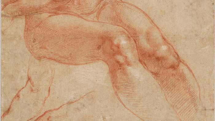
I am sure you have seen paintings and drawings by the old masters which depict the knee area so realistically that you have no doubts about its shape.
When drawing human figures from live memory or imagination, many art students have certain difficulties in depicting the knee joint. Some students oversimplify this part of the leg and this results in rather amature artwork.
To help you to avoid such junior mistakes, I created this video lesson.
Let us examine the knee anatomy.
The knee is a hinge-type joint. In this joint, the flat surface of the shinbone rotates around the cylindrical surface of the thighbone.
The main function of this joint is flexion and extension of the lower leg.
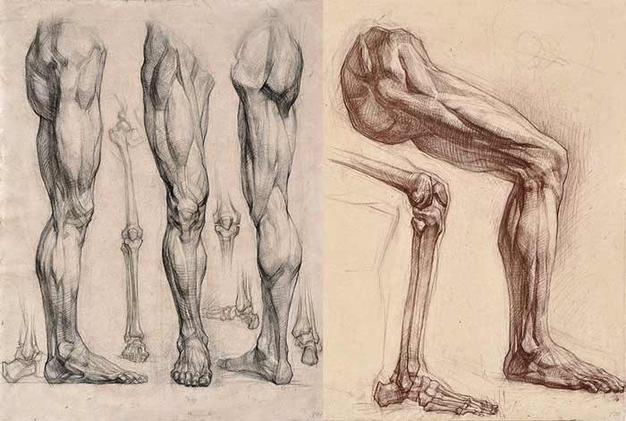
Knee Anatomy – Bones
The upper leg has one bone which is called the femur. The femur is the largest and heaviest bone in the human body. It is not straight, but slightly arched. This bone becomes wider at its lower end. This is to reduce the body weight pressure in the knee joint.
Two bony projections on either side of this bone are called the condyles of the femur.
In the front, just between two condyles, there is the knee cap which is called patella. Its name translates from Latin as small plate.
The patella, or the knee cap, has a triangular shape. Its upper edge coincides with the top of the condyles and its bottom edge is aligned with the lower end of the femur.
Another bone of the knee joint is called the tibia. This is the shinbone. The top part of the shinbone is twice wider than its shaft. This is to distribute the body weight and reduce pressure at the knee.
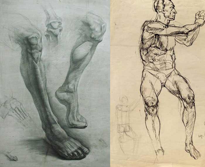
Underneath the kneecap, there is one bony projection the shinbone. It is called the tibial tuberosity. This is the place where muscle of the upper leg quadriceps connects to the lower leg bone. This tibial tuberosity plays an important role in the knee anatomy and must be depicted when drawing a leg.
At the side of the shinbone, there is another bone of the lower leg, which is called the calf bone, or the fibula. This bone is more slender than the tibia and does not participate in the knee joint.
Knee Anatomy – Muscles
A majority of the front portion of the upper leg is occupied by the four-headed muscle called quadriceps. It begins from the pelvis and the upper part of the thigh bone.
At the lower end, all four heads of this muscle come to a common tendon which encapsulates the knee cap and inserts into the tibial tuberosity of the shin bone just beneath the knee.
The tailor’s muscle, which spirals around the upper leg, goes from the pelvis and inserts into the inner condyle of the shin bone, not far from the knee joint.
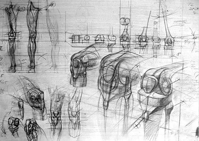
The width of the leg of the knee area does not depend upon muscles, but is actually defined by the dimensions of the bones.
The shin bone is detectable on the front surface of the leg. No muscles cover this bone here.
The calf muscle occupies the top-rear area of the lower leg. Its two heads are attached to both condyles of the thighbone. The main function of this muscle is to flex the lower leg at the knee joint.
Next time you draw a leg, do not forget about the knee anatomy. It includes the condyles of the femur and the condyles of the tibia as well as the knee cap and the tibial tuberosity…
[ The full lesson is avaibale to Anatomy Master Class members ]
To learn more about the knee anatomy, enroll in the Anatomy Master Class
Simple Pricing, No Surprises
One-time payment - Only $97 USD
ENROLL NOW