Human Muscles
Human Muscles
Anatomy Lesson 17 – Part 2
In this video lesson, you will discover the anatomy of human muscles.
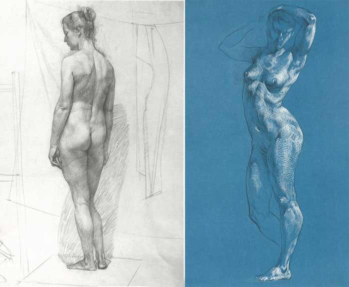
Human Muscles Anatomy for Figurative Artists
The deltoid muscle covers the shoulders from three sides- front, side, and back.
The biceps brachii muscle occupies the front portion of the upper arm. It is the main flexor of the forearm. The word biceps suggests this muscle has two heads. These heads originate from the shoulder blade.
The muscle of the neck also has two heads. One attaches to the top of the breastbone and another starts from the collarbone. They both insert into the base of the skull. Two neck muscles form the V-shape, with the pit of the neck between them.
On the back, the neck is covered by the upper portion of the trapezium muscle.
The chest muscle covers the frontal top part of the ribcage. It has three portions. The top one begins from the inner-half of the collarbone, the middle portion starts from the breastbone, and the lower portion begins from the sixth and seventh ribs. All three parts of this muscle insert into the upper arm bone.
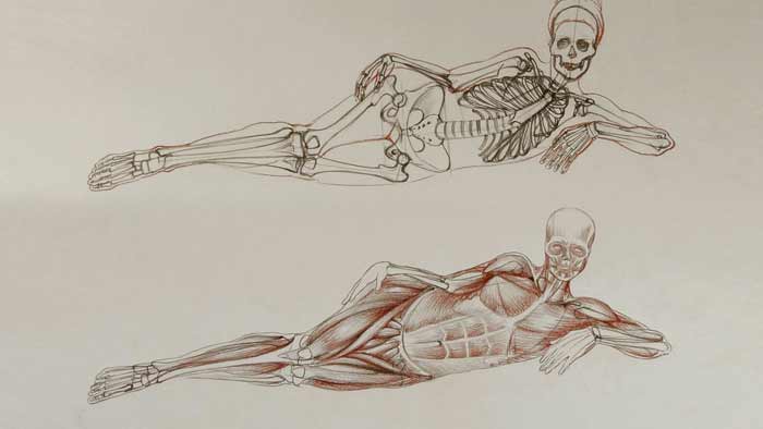
The frontal part of the abdomen is covered by the six-pack muscle. It begins from the pubic bone of the pelvis and inserts into the coastal cartilages of the fifth, sixth, and seventh ribs as well as into the lower part of the breastbone. Horizontally, it is separated by three white lines and vertically, another white line goes along its center. These lines divide the muscles into six-packs.
On both sides of the torso, there is a muscle, which begins from the first eight or nine ribs and inserts into the inner border of the shoulder blade beneath this bone.
The external oblique muscle covers the front and side parts of the abdomen. This muscle participates in many actions. It helps the six-pack bend the torso forward. It also flexes the torso sideways and rotates it.
Let us come back to the human muscles of the upper limbs.
The back portion of the upper arm is occupied by the triceps brachii muscle. It is the main extensor of the forearm.
The muscles of the forearm have elongated forms with long tendons. They can be divided into two groups – the flexors and the extensors.
The extensors muscle group is located on the posterior compartment of the forearm. Tendons of these muscles insert into various bones of the hand. These muscles extend and abducts the hand, thumb, and fingers.
The flexor group of muscles is located in the anterior compartment of the forearm. These muscles flex the hand and fingers.
Now, we will examine human muscles of the lower part of the body.
At the top of the pelvis, there is the muscle that raises the upper leg sideways. It begins from the upper edge of the pelvis and inserts into the top projection of the thighbone.
The tailor’s muscle starts from the top-front edge of the pelvis and travels diagonally downward. It inserts into the inner top part of the shinbone. It assist in pulling the leg in a cross-legged position.
The quadriceps muscle occupies the most area of the front part of the upper leg. It is responsible for flexing the thigh, at the hip joint, and extending the lower leg, at the knee. This muscle consists of four heads, which begin from the thighbone and the pelvis and insert, via the common tendon, into the knee cap and further down into the tibial tuberosity of the shinbone.
In the inner part of the upper leg, there is the adductor group of muscles. It consists of five muscles, which begin from the bones of the pelvis and insert into the thighbone. The main function of this group is to adduct the upper leg, or pull it closer to another leg.
In the front of the lower leg, there is an elongated muscle, which helps move the foot upward. Its tendon inserts into the middle of the foot and is easy to notice, at the lower front part of the leg, when the foot is bent upward.
Next to this muscle, there is another one, which goes alongside and inserts into the phalanges of the lesser toes. The primary function of this muscle is to extend the toes upward. It also helps raise the front part of the foot, at the ankle joint. Four tendons of this muscle are easy to spot on the surface of the foot when toes are raised upward.
There are two more muscles on the side of the lower leg, which begins from the calf bone and insert into the middle bottom part of the foot. These two muscles help move the foot downward, at the ankle joint.
The back compartment of the lower leg is occupied by the calf muscle and the soleus muscle. Together, they have three heads and share one common tendon, which inserts into the heel bone…
[ The full lesson is avaibale to Anatomy Master Class members ]
To learn more about human muscles every figurative fine artist must know, enroll in the Anatomy Master Class
Simple Pricing, No Surprises
One-time payment - Only $97 USD
ENROLL NOW