Head, Neck and Shoulders Skeletal Anatomy
Head, Neck and Shoulders Skeletal Anatomy
Anatomy Lesson 6 – Part 1
In this video lesson, you will discover the head, neck and shoulders skeletal anatomy.
How to Draw a Head and Shoulders Portrait – Skeletal Anatomy
The marble bust of the emperor Antonius Pius is used as a model for this drawing. This bust dates back to 140 A.D. and is displayed in the British Museum, London.
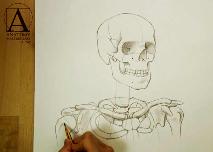
This video lesson begins with the drawing of a skull and explanation of its main proportions and features. This includes all major bones of the skull and their relative positions and proportions.
For example, the cheekbone is an important component of the cheeks, this bone also forms the outer and bottom portions of the eye sockets.
The dental area of the skull possesses a barrel shape.
The lower jaw is the only movable bone of the skull.
The upward projection of the mandible ends with two prongs.
The condylar process works like a hinge, touching the temporal bone. The jaw rotates around this joint.
The coronoid process resembles a shark fin. A powerful muscle that goes from the temporal fossa bone is attached to that process and, when contracted, lifts the lower jaw upward in a chewing movement.
The corner of the jawbone is called the angle of mandible. It is a distinctive mark every portrait artist needs to position correctly.
Dental arches vary from person to person and influence the surface of the cheeks and lips.
The upper dental arch is slightly bigger than the lower one.
Normally, a person has 32 teeth, which, depending on their location, shape, and function, can be divided into incisors, canines, premolars, and molars. The front teeth are called incisors; there are four of them on each upper and lower dental arch. Upper incisors are larger than lower ones.
The canine teeth follow the incisors and are shaped like cones. The premolars and molars succeed the canine teeth.
When drawing a portrait, it is advisable to avoid using a heavy outline on each tooth. A portrait will appear more natural when teeth are depicted in a more general way rather than hyper-realistically.
Also, do not forget to draw a cast shadow on the upper row of teeth under the upper lip. This will give depth to the mouth and allow the portrait to seem more natural.
In this video part, you will also find out the anatomy of the neck and shoulders.
The neck bones are called the cervical vertebrae. There are seven of them.
The first one that holds the skull is called the atlas.
The column of the neck bones is slightly curved.
The first pair of ribs resembles an oval and its size defines the neck width at its base.
The small axis of the first pair of ribs’ oval is perpendicular to the axis of the shoulders.
The outline of the ribcage travels down from the first pair of ribs.
The breastbone, or the sternum, follows the same perspective as the shoulder axis. The breastbone resembles a man’s tie and consists of three sections. There is an angle between the vertical axes of the top two sections. It is called the sternal angle. The top section of the sternum is similar to a knot of a tie and is called the manubrium of sternum.
Collarbones are attached to the manubrium of sternum.
Each collarbone has a twisted “S” shape. A collarbone begins from the pit of the neck, then travels, following the curve of the top rib. It then bends once more to follow the shoulder axis.
Together, two collarbones resemble a traditional bicycle handlebar.
The first rib is attached to the manubrium of sternum just below the collarbone. It does so via the costal cartilage.
The rib curves backward toward the spine and attaches to the first vertebra of the ribcage section.
In the front, the second rib attaches between the manubrium of sternum and the body of sternum. At the back, the second pair of ribs attaches to the second vertebra of the ribcage section of spine.
At the outer end, the collarbone meets the acromion of scapula, which is the outer part of the shoulder blade.
The upper arm bone, or the humerus, is connected to the shoulder blade.
The upper part of the arm bone is round like a golf ball and is called the head of humerus. This head sits inside of the joint that is a “ball and socket” connection. The socket, in this case, is the part of the shoulder blade called the glenoid fossa.
The bony projection of the shoulder blade is called the coracoid process. This process does not influence the shoulder’s appearance, but plays an important role for the shoulder muscles, which we will examine in following video sections.
Once again, let us list some anatomical parts of the shoulder region so you better understand its structure.
The collar bone connects with the acromion of the shoulder blade.
Other parts of the shoulder blade you must know are the coracoid process and the glenoid fossa (which is part of the shoulder joint).
The head of humerus, or upper arm bone, is another part of the shoulder joint.
[ The full lesson is avaibale to Anatomy Master Class members ]
To learn more about the head, neck and shoulders skeletal anatomy, enroll in the Anatomy Master Class
Simple Pricing, No Surprises
One-time payment - Only $97 USD
ENROLL NOW