Human Body Anatomy
Human Body Anatomy
Anatomy Lesson 20 – Part 2
In this video lesson, you will discover the human body anatomy on example of classical sculpture of David.
Human Body Anatomy for Figurative Artists
In the human body anatomy, the height of a standing human body can be divided in half. The mark in the middle locates the pubic bone of the pelvis.
In the contrapposto position, the pelvis is tilted; therefore, the spinal column is curved. The pelvis and the shoulders tilt in opposite directions.
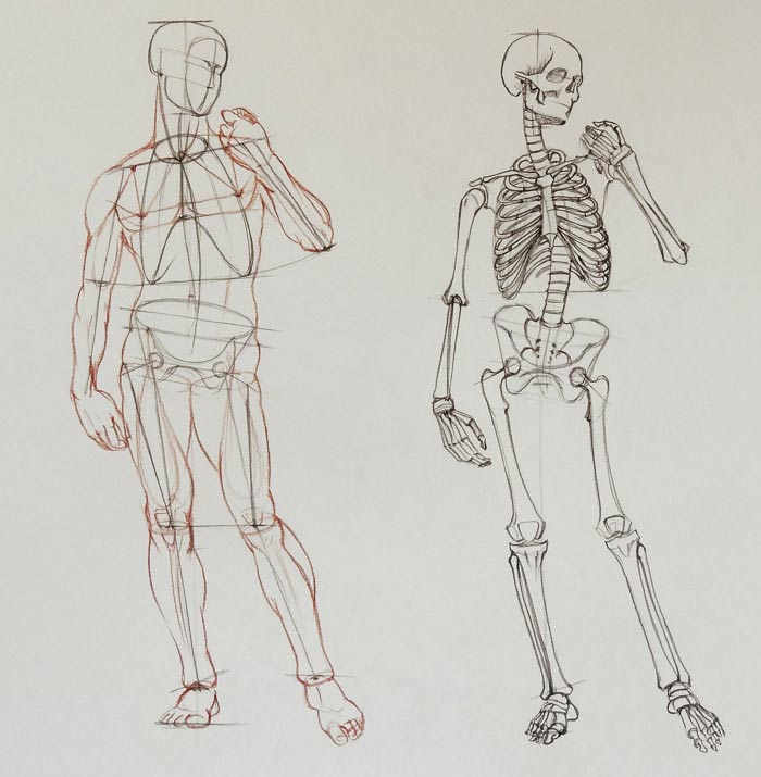
The spinal column is quite a complex structure; it consists of four regions: the neck, the rib cage, the waist, and the pelvic region. Each region has a specific curve when seen in profile.
The first pair of ribs is marked with an oval. Moving down from this pair, the ribcage grows steadily wider.
The breastbone is in the front. Its height is equal to the height of the face.
The pelvis is slightly wider than the ribcage. Its top edge is very close to the bottom of the ribcage. The pelvis consists of four different bones: the ilium, the sacrum, the sitting bones, and the pubic bone.
The thighbone connects to the pelvis via the ball-and-socket hip joint. There are two bony projections at the lower end of the thighbone. These projections are part of the knee joint.
The triangular kneecap protects the front side of this joint.
The shinbone is wider at the top; this creates a broad surface between the upper and lower leg bones, reducing the body’s pressure on the knee.
On the outer side of the shinbone is the calf bone.
The bottom projections of the shinbone and the calf bone are commonly known as the anklebones. The outer anklebone is lower than the inner one.
The bony bump on the frontal side of the shinbone just below the kneecap is called the Tibial Tuberosity. This is an important landmark and has to be depicted when drawing the knee.
The two collarbones follow the tilt of the shoulders. Their inner ends are attached to the top end of the breastbone.
The outer ends of the collarbones are connected to the shoulder blade.
The shoulder blade has two bony projections that I would like to mention here. These are the acromion and the coracoid processes. Several muscles attach to them.
The acromion sits above the round head of the upper arm bone, protecting it like a shield. This round head of the upper arm is part of the ball-and-socket shoulder joint. The socket of this joint is located on the shoulder blade.
The upper arm bone has two bony projections at the elbow.
The elbow bone of the lower arm is connected to the upper arm in a hinge-type joint.
The David’s right arm is in the pronation position. In this position the radius bone overlaps the elbow bone. In the anatomical position, these two bones are parallel to each other.
The hand consists of the wrist bones, metacarpal bones, and the phalanges of the thumb and fingers.
The David’s left arm is bent at the elbow.
The pointed tip of the elbow bone forms a triangle together with the two bony projections of the upper arm bone.
When drawing arms, it is important to understand the difference between the pronation and supination positions, as the direction of the forearm muscles will change with the direction of the radius as it rotates around the elbow bone.
The shoulder blade has a triangular shape. Its inner border is almost parallel to the spine when the shoulder blade is in its normal position.
The neck region of the spine has seven vertebrae. The ribcage region of the spinal column has twelve vertebrae.
The first pair of ribs is attached to the top of the breastbone, just below the collar bone.
The second pair of ribs is attached to the breastbone at the point where the head of the breastbone connects to its main body.
The third through seventh rib pairs are attached directly to the body of the breastbone.
Because the first seven pairs of ribs are connected to the breastbone, they are called the “true ribs.”
The next three pairs of ribs, from the eighth to the tenth, are not directly connected to the breastbone, but instead, each subsequent pair is attached to the previous one. That is why they are called the “false ribs.”
The last two pairs, the eleventh and the twelfth, are not connected at the front, and therefore are called the “floating ribs.”
At the back, every rib is connected to the vertebra at two points – to the body of the vertebra and to the process, which runs sideways from the vertebra.
The foot consists of seven tarsal bones, five metatarsal bones and fourteen phalanges (two in the big toes and three in each lesser toe).
More than half of all bones in a human body are located in the hands and feet…
[ The full lesson is avaibale to Anatomy Master Class members ]
To learn more about human body anatomy, enroll in the Anatomy Master Class
Simple Pricing, No Surprises
One-time payment - Only $97 USD
ENROLL NOW