Skull Anatomy
Skull Anatomy
Anatomy Lesson 18 – Part 1
In this video lesson, you will discover the skull anatomy.
Skull Anatomy for Portrait Artists
The model for this portrait will be the marble statue carved by French sculptor Jillian Christoff. This sculpture is displayed in the Victoria and Albert Museum in London.
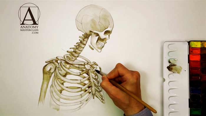
Let’s begin with the skull anatomy and the upper body skeleton.
The spinal column has two distinct curves- the curve of the neck region and the curve of the ribcage region. They are arched in the opposite directions.
At the top, there is the mass of the skull.
The upper part of the skull is called the cranium. It encapsulates and protects the brain.
The frontal lower side of the skull is the facial part.
We see the model’s head in profile. From this point of view, the head can be inscribed by a square.
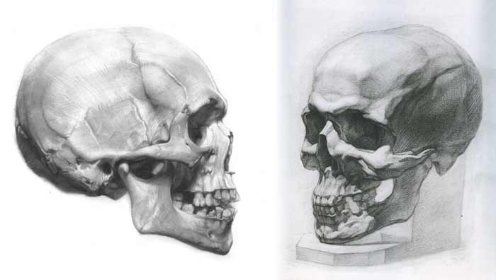
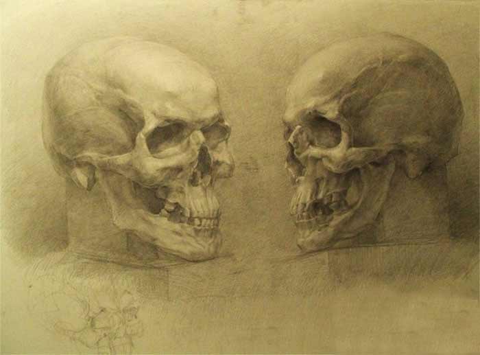
By dividing the height of the skull in half, we can locate the eye socket position.
The ear channel is located right above the first vertebra of the spine. Above it, there is the cheekbone arch.
The hinge of the lower jaw is located just in front of the ear channel underneath the cheekbone arch.
Right behind the ear channel, there is the bony process on the base of the skull. This is the place where neck muscle attaches to the skull.
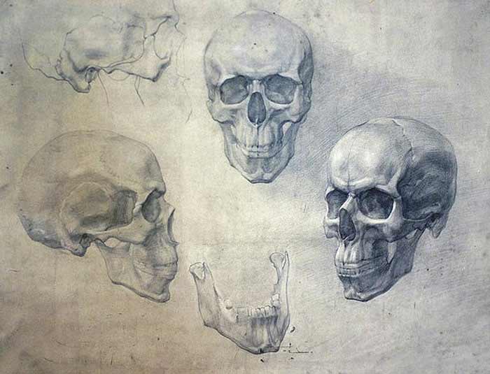
The cranium section of the skull consists of eight bones. They are fused together and forming the cranium dome and the cranium floor.
The facial part of the skull is formed by the fourteen bones.
The lower jawbone is the only bone of the skull that moves.
The U-shaped tongue bone is located underneath the lower jaw.
The neck region of the spine consists of seven vertebrae. This number is not unique for humans; in fact, many animals have seven bones in the neck spine.
The very top vertebra is called the atlas because it holds the sphere of the skull.
The joint between this vertebra and the skull allows the head rotate up and down in nodding movement.
This is the first pair of ribs. It has a circular outline and appears, in perspective, as an oval. The axis of this oval is slightly tilted downwards.
At the front, this pair of ribs is connected to the top part of the breastbone.
Just above the first rib, there is the collarbone. Its inner side is also attached to the top of the breastbone.
The collarbone is curved. The inner-half goes along the rib and the outer-half is bent to follow the shoulder axis.
At the back, the first pair of ribs is connected to the first vertebra of the ribcage region.
With every following rib, the ribcage becomes wider until it reaches the eighth rib where it’s the widest.
In the front, the second pair of ribs is connected to the breastbone in the place where the breastbone head and body meets together.
The frontal portion of every rib has softer and more flexible structure. It is called the cartilage.
There are seven true ribs, which means they are directly attached to the breastbone in the front.
The outer edge of the collarbone is connected to the shoulder blade in the part, which is called the acromion of the shoulder blade. This acromion hangs above the shoulder joint, protecting the head of the upper arm bone.
This head has a round shape of a size as a golf ball. Next to this round head, there is a bony projection, of the shoulder blade, which looks like a bird’s beak.
The upper arm has one bone. It is called the humerus. The groove next to the humerus head is the place for a muscle tendon…
[ The full lesson is avaibale to Anatomy Master Class members ]
To learn more about skull anatomy, enroll in the Anatomy Master Class
Simple Pricing, No Surprises
One-time payment - Only $97 USD
ENROLL NOW