Leg Anatomy
Leg Anatomy
Anatomy Lesson 12 – Part 4
In this video lesson, you will discover the leg anatomy.
Leg Anatomy – Bones
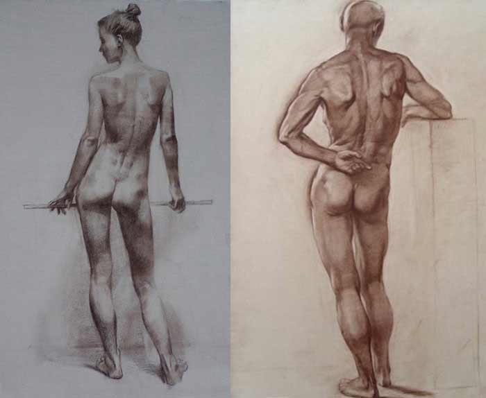
This video lesson will present you a drawing of the skeletal structure of the lower limbs in the contrapposto position. In this position, the pelvis’ main axis is not vertical, but tilted. The tilt of the pelvis occurs because it is supported by one leg. On the drawing below, the engaged right leg is straight while the left leg will be slightly bent at the knee.
The pelvis is seen from behind, which allows us to have a good view of the sacrum, ilium bones, and sitting bones.
The axis of the sockets of the hip joints is tilted together with the pelvis. It is located on the same level as the pubic bone.
The hip joint is a ball-and-socket type of joint. The ball, here, is the head of the thighbone, which is called the femur.
The neck of the femur is positioned diagonally downward in male anatomy.
In a female, these lines are more horizontal which, in general, make female hips a bit wider and legs shorter.
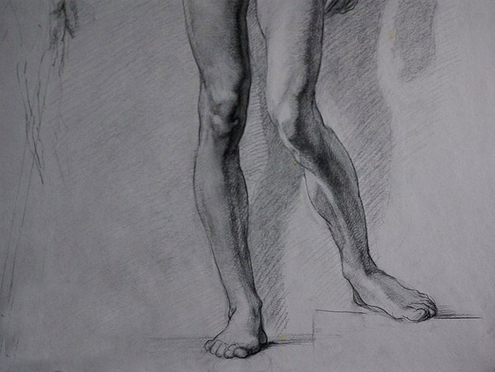
In the contrapposto position, the center of gravity of the human body vertically projects onto the footprint of the supporting leg so that the whole weight of the body is supported by just one leg. Another leg helps keep the body balanced.
The tilt of the thigh bone is not the same as the tilt of the shin bone. The axis of the shin bone points to the head of the femur while the angular positions of the femur’s neck and shaft make a long triangle.
Here is one important fact you, as a figurative artist, have to remember. The bottom ends of the shin bone and the calf bone are not on the same level.
The calf bone ends lower than the shin bone, so the ankle axis is tilted diagonally.
Once again, let us review the construction of the femur, or thigh bone. At the top, it starts with the head of the femur which has a ball shape.
Next, there is the neck of the femur, and two bony projections, the bigger and the smaller one. Many muscles attach to these projections.
The shaft of the thighbone is slightly bent forward. At the bottom end, this bone becomes wider and ends with two condyles (bony projections which form part of the knee joint).
There are two bones in the lower leg, the tibia, or shinbone and the fibula or calfbone.
Two condyles of the shinbone are part of the knee joint.
The calfbone bone does not participate in the joint and is attached to the outer side of the shinbone.
Leg Anatomy – Muscles
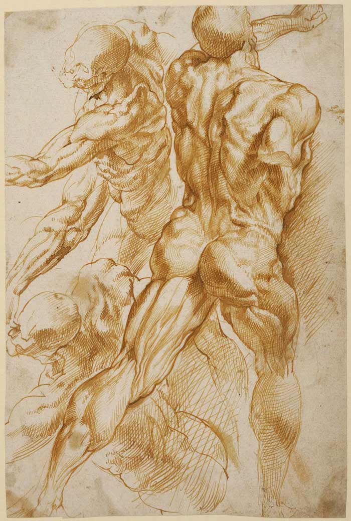
Now, it is time to examine the muscles of the lower limbs.
The gluteus medius muscle originates from the outer surface of the ilium bone of the pelvis and inserts into the top projection of the thighbone bone. This muscle helps move the upper leg away from the body and sideways.
Another muscle of the gluteal group is the gluteus maximus. It goes from the back edge of the pelvis and inserts into the upper part of the femur as well as, via a long and flat tendon, into the top outer side of the shinbone. This powerful muscle moves the thigh backward in extension of the lower limb. Gluteal group muscles belong to the muscles of the torso.
The hamstring group of muscles is located on the back of the thigh. It resembles a cylindrical mass and covers most of the rear portion of the upper leg. The hamstring group consists of three muscles. They all originate from the sitting bone of the pelvis. The hamstring muscles insert, via tendons, into both sides of the shinbone and calfbone, below the knee joint. The triangular pit between these muscles is called the ham. That is where the name of the hamstring muscles originates from.
As I mentioned, there are three muscles in that group. Two muscles overlap each other and travel downward to the inner side of the knee and there is one more muscle that goes to the outer side. Hamstring muscles tendoins can be seen at the back of the knees.
There are five muscles that form the adductor group. Adductor group of muscles begins from the sitting bone and the pubic bone and inserts into the thighbone. One muscle inserts into the tibia. Together, these muscles form the inner portion of the thigh.
As indicated by its name, the adductor group of muscles has the primary action of adducting the upper leg at the hip joint. Some muscles of this group also contribute to the flexion and extension of the hip.
Now, let’s examine the muscles of the lower leg.
Most of the back side of the lower leg is occupied by the calf muscle. It possesses two heads (an outer and inner one). These two heads originate from points just above two condyles at the lower rear side of the thigbone. At the bottom, they share the common tendon which inserts into the heel bone. This tendon is referred to as the Achilles tendon. This muscle assists in the flexion of the lower leg at the knee. It also takes part in flexing the foot downward as well as raising the heel from the ground when standing tip-toe.
Underneath the calf muscle, there is one more muscle called the soleus. This muscle also attaches to the heel bone. Its name comes from the Latin word fish which refers to the shape of this muscle. This muscle engages in similar actions as the calf muscle…
[ The full lesson is avaibale to Anatomy Master Class members ]
To learn more about the leg anatomy, enroll in the Anatomy Master Class
Simple Pricing, No Surprises
One-time payment - Only $97 USD
ENROLL NOW