Female Body Anatomy
Female Body Anatomy
Anatomy Lesson 16 – Part 2
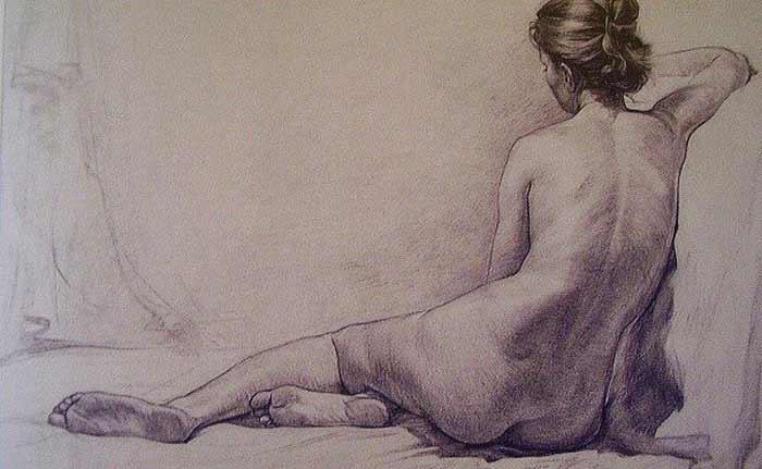
In this video lesson, you will discover the female body anatomy.
Female Body Anatomy for Figurative Artists
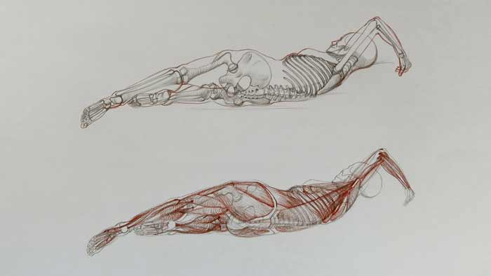
Let’s begin with the muscle anatomy of female body.
The deltoid muscle rear portion starts from the the shoulder blade and inserts into the middle of the upper arm bone shaft.
The front part of the upper arm is occupied by the biceps brachii. It is the principle flexor of the arm.
At the rear side of the upper arm, there is a muscle called the triceps brachii. It is the main extensor of the forearm. It inserts into the tip of the elbow bone.
From the outer epicondyle of the upper arm bone starts a group of forearm muscles, which is the extensor group of the lower arm. There are four muscles in this group. Three of them share the common tendon, which starts from the outer epicondyle.
These extensor muscles insert into the various bones of the hand. Their main function is to extend the wrist and fingers, straightening them from a bent position.
The chest muscle arises from the inner-half of the collarbone, coastal cartilages of true ribs and the breastbone, and the sixth anf the seventh ribs. This muscle inserts into the upper arm bone.
Another muscle of the torso, which we have a good view of in this position, is called the serratus anterior. It begins from the outer surfaces, of the upper eight or nine ribs, and is located on the side of the ribcage. This muscle inserts into the inner border of the shoulder blade. Since it travels along the ribs, it is, sometimes, mistaken on the surface for ribs on the side of the chest.
There are several muscles between the shoulder blade and the upper arm bone. The muscle, which goes from the lower end of the shoulder blade and inserts into the upper arm bone is called the teres major. This muscle adducts the arm, pulling it towards the torso, and also rotates it inward and extends it backward.
Close to this muscle, there is another muscle called the teres minor. On the surface of the torso, it is difficult to detect this muscle because it is partly covered by and blends with another muscle, the infraspinatus. This muscle rotates the arm outward.
The widest muscle of the back is the latissimus dorsi. It covers a large portion of the back. It assists in three movements of the arm:
- First, it extends the arm, pulling it from the raised position in front of the body, toward the torso.
- Second, it adducts the arm, pulling the arm sideways towards the torso.
- And lastly, it assists in medial rotation – rotation of the arm inward.
The external oblique muscle covers the front and side portions of the abdomen. Here, we see its lower side portion.
The gluteus medius muscle is between the top border of the pelvis and the upper edge of the thighbone.
When drawing a live figure, do not confuse this muscle with the flank pads of the external oblique muscle.
The flank pads are above the iliac crest of the pelvis and the gluteus medius is below.
The buttocks muscle, or the gluteus maximus, is a well-pronounced muscle, which starts from the rear part of the pelvis and inserts into the outer top part of the thigh bone. It also inserts into the top of the shinbone via the flat long tendon. This tendon is also the place of insertion of the third muscle of the gluteal group.
The back portion of the thigh is occupied by the hamstring group of muscles. This group consists of three muscles. These muscles begin from the sitting bone and one muscle, of this group, originates from the thighbone.
They insert into the upper part of the lower leg bones. The main action of these muscles is to extend the thigh.
The rear part of the lower leg is occupied by the calf muscle and the soleus muscle. They insert, via the common tendon, into the heel bone.
Two heads of the calf muscle begin from the outer and inner condyles of the thighbone. These muscles flex the lower leg and move the foot downward.
On the side of the lower leg, there is a muscle that starts from the calf bone and inserts into the foot bones. It helps move the foot downward.
Next to it, there is another muscle which has the same function; however, it inserts into the fifth metatarsal bone of the foot…
[ The full lesson is avaibale to Anatomy Master Class members ]
To learn more about female body anatomy, enroll in the Anatomy Master Class
Simple Pricing, No Surprises
One-time payment - Only $97 USD
ENROLL NOW