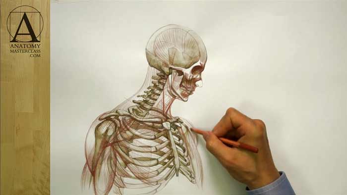
Facial Anatomy
Facial Anatomy
Anatomy Lesson 18 – Part 2
In this video lesson, you will discover the facial anatomy every figurative fine artist must know.
Facial Anatomy for Portrait Artists
Let’s begin with muscles of the human head.
The muscle of the forehead extends from the hairline to the brow ridge. It is actually the frontal portion of a much bigger muscle that spans from the brow ridge to the back edge of the skull.
Another portion of this muscle is located on the back of the head. Between these two portions there is wide flat tendon, which covers the entire top part of the skull.
The forehead muscle can produce different expressions on a face. Contraction of both left and right portions of this muscle elevates the eyebrows, which gives an impression of surprise or disbelief. When only one side is contracted, one eyebrow is lifted. This arching of the eyebrow expresses bemusement or distrust. In fact, not every person can produce such action of the forehead muscle. This muscle can also lift upward the inner corners of eyebrows, making an expression of sadness or grief.
The sidewalls of the skull are covered by flat, fan-shaped muscle, which inserts into the process of the lower jawbone. This muscle is closing the jaw and retracts it.
There is another muscle that mainly is responsible for closing the jaw. It is located on both sides of the lower jawbone. This muscle is easy to spot when a person clenches the teeth or chews something hard. It can indicate the emotional tension.
There are three portions around the ear channel of the muscle, which is actually obsolete as human lost the ability to move their ears in the sound direction.
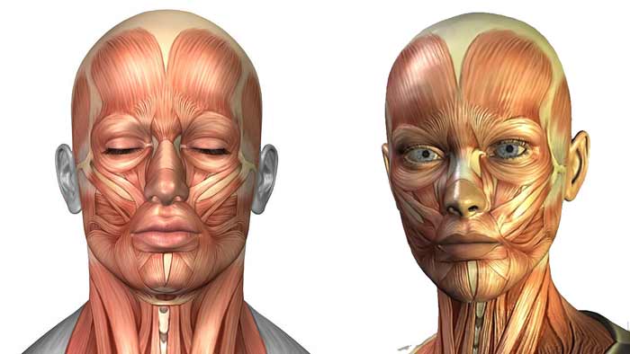
Around the eyeball there is the disc-shaped muscle. It has two parts: the thicker part that goes around the eye and the slimmer one, which is located inside the eyelids. The thicker fibers of the circular eye muscle squeeze the eye shut. This muscle is in action when someone squints or winks. The slender, and more delicate, portion in the eyelids gently closes eyelids when blinking.
There is another important muscle of the human head, which has a circular shape. This is the muscle of the mouth. This muscle closes the lips, pulling them together.
The trumpeter’s muscle goes horizontally sideways from the corner of the mouth. This muscle pulls angle of the mouth straight back, compressing cheeks and lips. It is used when a person is blowing a trumpet. That’s where its common name comes from.
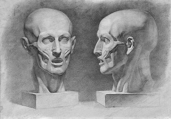
There are four muscles on either side of the face that begin from the cheekbone and insert into the upper part of the mouth muscle.
The muscle, which is the closest to the nose, elevates the wings of the nose and pulls upward the middle portion of the upper lip.
Three other muscles are also pulling upward the upper lip as well as stretching it out. Working separately and together, they can produce various expressions on a human face. They can produce a wide array of emotions from disgust to smiling.
On either side of the nose, there is the nose muscle, which is quite difficult to detect. It compresses the nostrils and pulls the nose slightly downward, which slightly enlarges the nostrils.
Near the root of the nose there is a couple of muscles which depress the inner ends of the eyebrows, producing horizontal wrinkles near the root of the nose. It displays an expression of annoyance, anger, or intense concentration.
In the facial anatomy, there is the muscle which pulls the corners of the mouth downward. It participates in expressions of sadness of disapproval.
Next to it, there is another muscle that begins on the lower jaw and inserts into the lower lip. When contracted, it pulls the lower lip downward. It is an important muscle as it participates in speech.
And finally, there is one more muscle in the chin region. When contracted, it pulls the skin of the chin upward. It can participate in various expressions like anger, sadness, or determination.
Underneath the lower jaw, there is a couple of muscles, which connects the lower jaw, the tongue bone, and the base of the skull. These muscles help to open the lower jaw as well as to elevate the tongue bone when swallowing and speaking.
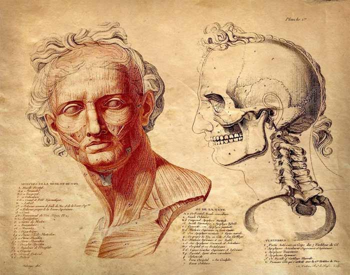
The front portion of the neck has the Adam’s apple, cartilage, and the windpipe. This structure is also known as the voice box.
Here is the top portion of the back muscle, which, among other bones, also inserts into the base of the skull.
The muscle of the neck has two heads. One begins from the top edge of the breastbone and another from the inner part of the collarbone. These two portions insert into the bony process and base of the skull, behind the ear channel. Working together, these neck muscles bend the head into different positions- forward, backward, and sideways, as well as rotating it from left to right.
The trapezium muscle covers the rear side of the neck. This diamond-shaped muscle begins from the base of the skull and vertebrae of the spine and inserts into the outer part of the collarbones and spines of the shoulder blades.
The front of the neck is covered by thin, flat muscle that is located between the collarbones and the lower jaw. There is a muscle, which connects the ribs between each other.
On the side of the torso, there is the muscle, which has eight or nine digitations that start from side parts of the ribs and insert into the inner border of the shoulder blade. The main function of this muscle is to move the shoulder blade.
The biceps brachii is an important muscle of the upper arm. As indicated by its name, it has two heads that begin from the shoulder blade. The tendon, of one head, goes along the groove on the upper arm bone. This muscle occupies the entire front portion of the upper arm. Its main action is to flex the forearm.
The back portion of the upper arm is occupied by the triceps brachii, which extends the forearm.
The chest muscle has three portions. The top portion begins from the inner half of the collarbone and travels diagonally downward. The middle portion begins from the breastbone and travels horizontally sideways. And the third lower portion begins from the sixth and seventh ribs and travels diagonally upward. These three portions insert into the upper part of the upper arm bone. Fibers of this muscle overlap each other near the place of insertion. So the point of insertion of the upper portion is lower than the point of insertion of the lower portion.
The chest muscle moves the upper arm bone. It participates in the actions of adduction, flexion, and medial rotation.
Another important muscle of the shoulder girdle is the deltoid. This muscle consists of three parts – the frontal portion, the side portion, and the back portion. It begins in front from the outer half of the collarbone, the side portion begins from the outer edge of the shoulder blade, and the back portion begins from the shoulder blade spine.
The deltoid overlaps the chest muscle and inserts approximately in the middle of the upper arm bone. It has triangular shape. It assists in the actions of raising the arm horizontally (which is abduction) and raising the arm out in front of the body (which is flexion). It is also contributes to the action of moving the arm back, behind the torso (which is extension)…
[ The full lesson is avaibale to Anatomy Master Class members ]
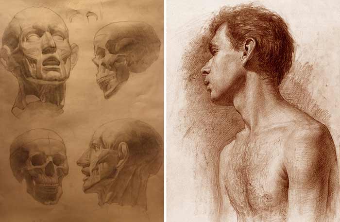
To learn more about facial anatomy and muscles of the upper body that every portrait artist must know, enroll in the Anatomy Master Class
Simple Pricing, No Surprises
One-time payment - Only $97 USD
ENROLL NOW