Body Anatomy
Body Anatomy
Anatomy Lesson 16 – Part 1
In this video lesson, you will discover the body anatomy on an example of drawing a reclining female figure.
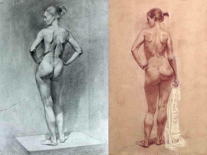
Body Anatomy for Figurative Artists
Let’s begin with the Skeletal Anatomy.
The spine has characteristic arches at the neck, ribcage, waist, and pelvis regions. Every region is arched into a position that is opposite to the previous one.
The lumbar region of the spine, which is in the waist area, has five vertebrae.
Above this region, there are two more sections of the spine, which are the cervical region in the neck area and the region of the ribcage.
The ribcage region of the spinal column consists of twelve vertebrae.
The neck region of the spine consists of seven vertebrae.
The first vertebra is called the atlas. The skull connects to this bone.
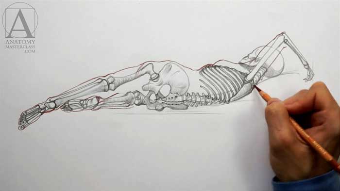

The sacrum is positioned between two ilium bones of the pelvis. The sacrum is the central structure of the pelvis. It consists of five vertebrae fused together into one triangular bone.
At the top, the ribcage begins with the first pair of ribs. The breastbone marks the front edge of the ribcage.
The rib pairs from the first to the seventh are true ribs.
The eighth, nineth, and tenth ribs are false ribs. This comes from the fact that they are not attached to the breastbone directly, but instead, in the front, they are connected to the coastal cartilage of the previous rib.
The last two ribs are only connected to the spine. Their front ends are floating unattached. This is why these two ribs are called floating ribs.
The bone of the upper arm is called the humerus. It is connected to the shoulderblade via the shoulder joint.
The elbow bone is wider at the elbow joint and is narrower at the wrist joint.
Another bone of the lower arm is the radius. This bone is wider at the wrist.
The carpal bones form the wrist and the metacarpal bones are part of the hand-block.
In female body anatomy, the neck of the thighbone is positioned almost at a right angle (not diagonally like in male anatomy). Therefore, female hips are slightly wider than male hips.
The larger bone of the lower leg is the shinbone. The upper edge of this bone is also a part of the knee joint. The lower edge of the shinbone is part of the ankle joint. Its inner bony projection is the inner anklebone.
Another bone of the lower leg is called the calf bone. At its bottom edge, it is positioned lower than the shinbone. This is why the outer anklebone is lower than the inner one. At the top, the calf bone is connected to the shinbone. The shaft of the calf bone is slimmer than the shaft of the shinbone…
[ The full lesson is avaibale to Anatomy Master Class members ]
To learn more about body anatomy, enroll in the Anatomy Master Class
Simple Pricing, No Surprises
One-time payment - Only $97 USD
ENROLL NOW