Anatomy of the Leg
Anatomy of the Leg
Anatomy Lesson 12 – Part 3
In this video lesson, you will discover the Anatomy of the Leg for figurative artists.
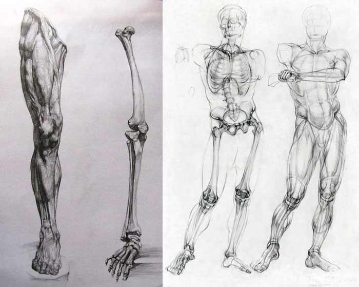
Anatomy of the leg – Bones
Let’s begin with the skeletal anatomy.
Anatomically, the term leg refers only to the lower leg. The term lower limb indicates the upper and the lower leg (from the hip joint to the ankle joint).
The femur is the bone of the upper leg. It is also called the thigh bone. This is the largest bone in the human body. It has the following structure. At the very top, there is a head. This head is part of the ball and socket hip joint. The head of femut is connected to the bone’s shaft via the neck. There are two bony projections next to the neck, the bigger on the top and the smaller one below. The shaft of the femur is slightly bent forward. At the bottom, this bone becomes wider and ends with two bony projections on the inner and outer sides. These projections are called condyles, the inner condyle and the outer condyle of the femur.
The lower leg has two bones. The larger one is called the tibia, or the shin bone. This bone is also wider at the knee joint. Two bony projections of the tibia are also called the condyles. The tibial tuberosity is located beneath the knee cap.
On the outer side of the tibia, there is the bone called the fibula, or the calf bone.
When drawing anatomy of the leg, keep in mind that the lower end of the fibula is lower than the bottom edge of the tibia. This is important to remember when depicting ankles.
The upper projection of the femur is located close to the body surface. You can actually feel this bone when touching your hips.
At the knee area, the width of the leg is defined by the dimensions of the condyles of the femur and the tibia.
The width of the ankle is also defined by the bony ends of the tibia and fibula.
The upper leg bone, the femur, is surrounded by muscles on all sides.
This is not the case for the tibia. This bone is quite close to the leg surface at the front. You can actually feel this bone when touching the front surface of the lower leg.
The tibial tuberosity and the knee cap are noticeable landmarks of the knee area.
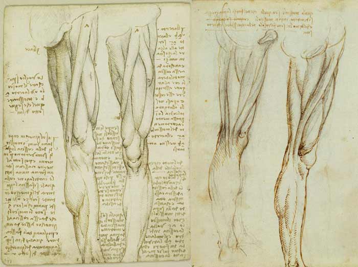
Anatomy of the Leg – Muscles
Let’s examine the muscles of the upper and lower leg.
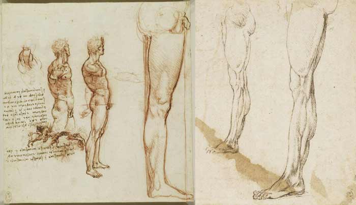
In the front portion of the upper leg, there is a muscle called the Sartorius. Sartor in Greek means tailor. This is why this muscle is also referred to as the tailor’s muscle. The tailor’s muscle starts from the frontal upper edge of the pelvis and inserts into the inner top portion of the shinbone. Its main function is to help other muscles to bring the leg into the crossed-leg position. The tailor’s muscle is long and flat like a belt. It goes diagonally downward and inward around the upper leg.
There is another leg muscle that goes outward from the same point of the pelvis. Via a flat tendon on the outer side of the upper leg, it inserts into the upper outer side of the shinbone. This muscle helps move the lower limb outward and sideways.
Another muscle that contributes to this movement is attached to the iliac crest, or the upper edge of the pelvis, and inserts into the top edge of the thighbone. This muscle is called gluteus medius.
Most of the front portion of the upper leg is occupied by the quadriceps group of muscles. As suggested by its name, this group has four heads. One is attached to the pelvis and three other heads originate from the top part of the femur. All four heads of this group come to one tendon which encapsulates the knee cap and inserts into the tibial tuberosity of the shinbone. The quadriceps is a very powerful group of muscles. Its primary function is to extend or staighten the leg at the knee. You can feel how it works when climbing upstairs.
Although the quadriceps group of muscles has four heads, only three of those can be detected on the surface of the thigh. Usually, these three heads are seen as the unified mass of muscles.
There are five muscles that form the adductor group. This group is located in the inner upper portion of the thigh.
The two headed calf muscle occupies the upper rear half of the lower leg. This muscle goes from the lower part of the femur and inserts, via a tendon, into the heel bone. Its main action is to bend the lower leg at the knee joint and flex the foot downward.
The shin muscle travels diagonally downward on the lower leg and helps to lift the foot when walking.
On the outer side of the lower leg, there is one more muscle that goes from the top outer side of the shin bone and inserts, via four tendons, into the toe phalanges of the foot from the second to the fifth toes.
Underneath the calf muscle, there is the soleus muscle which helps flex the foot downward and stand on the tips of the toes…
[ The full lesson is avaibale to Anatomy Master Class members ]
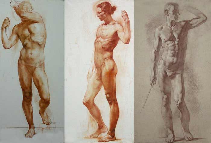
To learn more about the anatomy of the leg, enroll in the Anatomy Master Class
Simple Pricing, No Surprises
One-time payment - Only $97 USD
ENROLL NOW